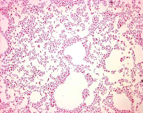
Bacillus megaterium is a gram-positive bacterium, meaning it retains the crystal violet stain during the Gram staining procedure. When subjected to the Gram staining technique, B. megaterium will appear purple under the microscope due to its thick peptidoglycan layer in the cell wall, which retains the crystal violet stain after the decolorization step.
How to Perform Gram Staining on Bacillus megaterium?
Gram staining is a widely used microbiological technique to differentiate bacteria into two groups based on their cell wall composition: Gram-positive and Gram-negative. Bacillus megaterium is a Gram-positive bacterium, meaning it has a thick peptidoglycan layer in its cell wall. Here’s a basic protocol for performing Gram staining on Bacillus megaterium:
Materials
- Bunsen burner or alcohol lamp
- Microscope slides
- Bacterial culture of Bacillus megaterium
- Crystal violet (primary stain)
- Gram’s iodine (mordant)
- Decolorizer (e.g., ethanol or acetone)
- Safranin (counterstain)
- Distilled water
- Microscope
- Immersion oil (if using an oil immersion objective)
Procedure
Prepare a clean microscope slide by labeling it with the name of the sample.
Take a small loopful of Bacillus megaterium culture using a sterile inoculating loop and spread it thinly over the surface of the slide to create a bacterial smear. Let it air dry.
Fix the bacterial smear by passing the slide, bacterial side up, through the flame of a Bunsen burner or alcohol lamp several times. Be careful not to overheat the slide.
Flood the bacterial smear with crystal violet stain and let it stand for 1 minute. Crystal violet will stain all cells purple.
Rinse the slide gently with distilled water to remove excess crystal violet.
Flood the slide with Gram’s iodine (iodine solution) and let it stand for 1 minute. Gram’s iodine functions as a mordant, forming a complex with crystal violet and enhancing its retention within the bacterial cells.
Rinse the slide gently with distilled water to remove excess iodine.
Decolorize the slide by gently flooding it with decolorizer (ethanol or acetone) drop by drop until the runoff is clear (usually 10-20 seconds). Be cautious not to over-decolorize, as this can lead to false results.
Rinse the slide immediately with distilled water to stop the decolorization process. Counterstain the smear with safranin for 1 minute. Safranin will stain Gram-negative cells pink, but it will have little effect on Gram-positive cells that have retained the crystal violet-iodine complex.
Rinse the slide gently with distilled water to remove excess safranin.
Blot the slide gently with bibulous paper or allow it to air dry.
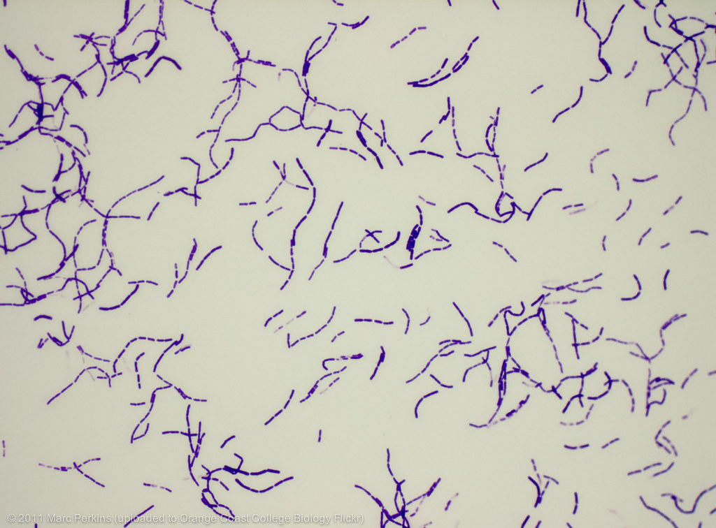
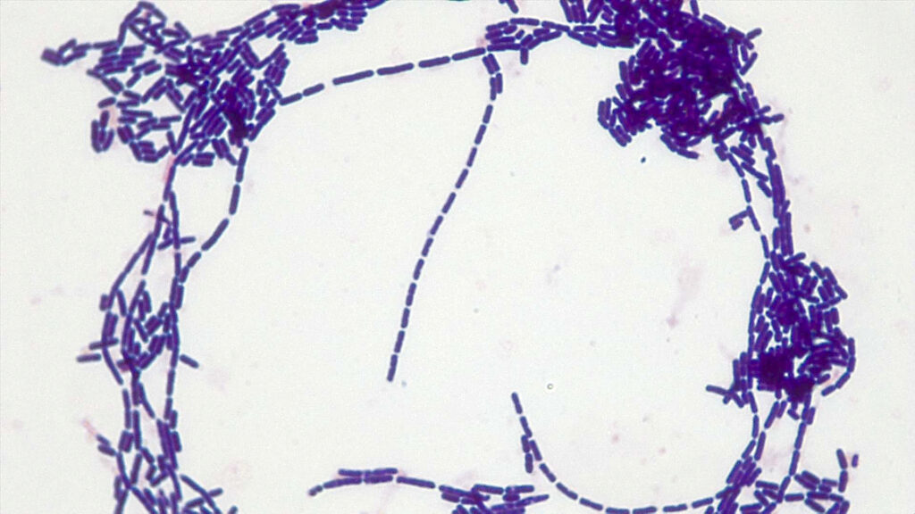
Examine the slide under a microscope using the oil immersion objective if available. Bacillus megaterium cells should appear purple, indicating that they are Gram-positive.
What Are the Gram Staining Results for Bacillus megaterium?
When performing Gram staining on Bacillus megaterium, the thick peptidoglycan layer in its cell wall plays a crucial role in determining its Gram staining results.
After the initial application of the primary stain, crystal violet, Bacillus megaterium cells will take up this stain and appear purple. The crystal violet molecules penetrate through the thin layer of peptidoglycan in Gram-negative bacteria but are trapped within the thick peptidoglycan layer of Gram-positive bacteria like Bacillus megaterium.
Following the application of the mordant, Gram’s iodine, which forms a complex with the crystal violet, the purple color intensifies. This complex is not easily removed from the thick peptidoglycan layer of Gram-positive bacteria during the subsequent decolorization step.
During the decolorization step, the alcohol or acetone removes the lipid-rich outer membrane of Gram-negative bacteria, allowing the crystal violet-iodine complex to be washed away, rendering them colorless. However, the thick peptidoglycan layer of Gram-positive bacteria like Bacillus megaterium acts as a barrier, preventing the removal of the crystal violet-iodine complex, thus retaining the purple color.
Finally, when the counterstain, safranin, is applied, it does not significantly alter the appearance of Bacillus megaterium cells because they have already retained the purple color from the crystal violet-iodine complex. Thus, under the microscope, Bacillus megaterium cells will appear purple, confirming their classification as Gram-positive bacteria.
Final Words
Gram staining is a vital technique in microbiology for identifying bacteria like Bacillus megaterium. By following a simple procedure, scientists can determine whether the bacterium is gram-positive or gram-negative based on its reaction to stains. This method helps in bacterial classification and has important applications in both clinical and research settings. Understanding the significance of Gram staining enhances our knowledge of B. megaterium and its role in various fields, from medicine to industry.